
3D / 4D Pregnancy Ultrasound (Sonography) in Ahmedabad
A pregnancy scan is the moment most parents look forward to, and also the one they quietly dread. Understanding what each scan is actually for takes some of that anxiety away.
Different scans answer different questions - dating, screening, detailed anatomy, growth, blood flow. Each is done at a specific point for a specific reason.
At Divine Women's Hospital, Ahmedabad, scanning is performed and interpreted in-house by Dr. Devang Patel, who holds Nuchal Translucency accreditation from the Fetal Medicine Foundation, UK.

Reports are only as good as the person holding the probe. Every scan here is performed and interpreted by experienced hands, and explained to you on the screen before you leave.

Overview of Ultrasound
Diagnostic ultrasounds use sound waves to create images of the body. Ultrasound also known as sonography shows the arrangements inside the body. The pictures can assist guide diagnosis & treatment for many diseases & disorders.
Most ultrasounds are performed using an instrument outside the body. Yet, some apply to place a small instrument inside the body.
Why is Ultrasound done?
Ultrasound is performed for many bases, including:
- To View the uterus & ovaries during pregnancy & to monitor the newborn baby’s health.
Analyze gallbladder condition. - Estimate blood flow.
- Guide a needle for tumour treatment.
- Read a breast lump.
- Review the thyroid gland.
- See genital & prostate problems.
- Consider joint inflammation called synovitis.
- Consider metabolic bone disorder.
Types of Ultrasound
During fetal check-ups, many types of ultrasounds are performed to evaluate the fetus’s development and health. Each category serves a unique purpose and offers different levels of detail. The following are the most common types of ultrasounds used during fetal examinations:
Trans-abdominal ultrasound:
This is the most common form of ultrasonography used in pregnancy. A transducer is moved around the abdomen to produce images of the fetus and surrounding structures. It is used for routine checks to monitor fetal growth, detect multiple pregnancies, and determine the position of the placenta.
Transvaginal ultrasound:
This method involves inserting a tiny transducer into the vagina to provide more precise pictures of the cervix and lower uterus. It is commonly used in early pregnancy to confirm pregnancy, monitor fetal heartbeat, and assess early fetal development.
3D Ultrasound:
This approach generates three-dimensional photographs of the fetus, allowing for more detailed views of its anatomy. It is especially beneficial for examining fetal anatomy like the face, limbs, and organs.
4D Ultrasound:
Similar to 3D ultrasound, but with the addition of time, resulting in live, moving photos of the fetus. It allows doctors and parents to watch the fetus’ movements and expressions in real time.
Doppler Ultrasound:
Doppler Ultrasound assesses blood flow in the umbilical cord, placenta, and fetal arteries. It is used to assess fetal health, particularly in cases of suspected fetal growth restriction or placental insufficiency.
Fetal echocardiography:
A specialist ultrasound for studying the fetal heart. It offers detailed views of the heart’s anatomy and function, making it easier to discover congenital heart abnormalities.
Anatomy Scans (Level II Ultrasound):
This thorough scan, which is typically performed between 18 and 22 weeks of pregnancy, examines fetal anatomy for anomalies and assesses organ and limb development.
The biophysical profile (BPP):
To measure fetal health, this test combines an ultrasound with a non-stress test. It assesses fetal movements, muscle tone, breathing patterns, and amniotic fluid volumes.
Nuchal Translucency Scan:
This ultrasound is performed between 11 and 14 weeks and assesses the thickness of the fluid at the back of the fetus’ neck. It is used in conjunction with blood testing to screen for chromosomal abnormalities such as Down syndrome.
Each type of ultrasound gives significant information to healthcare providers, allowing them to monitor the fetus’s health and make informed pregnancy management decisions.
Ultrasound is a safe, non-invasive imaging method that can reveal important information about the mother’s and baby’s health and growth throughout pregnancy.
Divine Women’s Hospital provides treatment and services to women, particularly in obstetrics and gynecology, including ultrasounds to pregnant mothers.
Dr. Devang Patel is an obstetrician at Divine Women’s Hospital who performs pregnancy ultrasounds, with a commitment to patient care and precision in prenatal imaging.
Ultrasound treatment cost in Ahmedabad
What treatment costs depends on the service required, how many visits or scans are involved and the complexity of your case.
| Treatment | Estimated cost range |
|---|---|
| Consultation and routine checks | ₹500 – ₹1,500 per visit |
| Scans and diagnostic tests | ₹2,000 – ₹7,000 |
| Medical management | ₹10,000 – ₹50,000 per year |
| Procedure or delivery package | ₹50,000 – ₹1,50,000+ |
These are indicative ranges rather than a quotation. Consultations, scans, medication and hospital stay vary with your plan - you are given a written estimate for your own care after your consultation.
Why Choose Divine Women's Hospital for Pregnancy Ultrasound
- Nuchal Translucency accreditation from the Fetal Medicine Foundation, UK
- Scans performed and interpreted by your own consultant, not sent out for reporting
- 3D and 4D imaging, Doppler studies and fetal echocardiography available
- In-house fetal medicine assessment where a scan raises a question
- Results explained to you at the time rather than left to a report you cannot read
- Scans grouped with consultations where you are travelling from out of town
Pregnancy Ultrasound Specialist in Ahmedabad

At Divine Women's Hospital, Ahmedabad, the person scanning you is the person interpreting the images and planning your care - which is why findings are explained at the time rather than in a letter.
Dr. Devang Patel holds an M.D. in Obstetrics & Gynecology and an FNB in High Risk Pregnancy and Perinatology, and is accredited for Nuchal Translucency scanning by the Fetal Medicine Foundation, UK.
Scanning, fetal medicine assessment, theatre and NICU support are all on site.
A scan is only as good as the person reading it - and the conversation that follows.
If a scan is due, or one has raised a question you want explained properly, book an appointment.
Frequently Asked Questions
Yes. Ultrasound uses sound waves rather than radiation and has been used in pregnancy for decades with no evidence of harm. Scans are performed when there is a clinical reason.
What to expect at Divine Women's Hospital
Choosing where to be cared for is a personal decision, and it usually comes down to one question: will someone actually listen to me? At Divine Women's Hospital, that is where every consultation begins. Our senior consultants stay with you from the first visit through recovery, so you never have to repeat your story to a new face.
One consultant, start to finish
The doctor who examines you is the doctor who plans your treatment and performs your procedure. Continuity is not a luxury here, it is the baseline.
Explained in plain language
Reports are walked through line by line, in Gujarati, Hindi or English, until the plan makes sense to you and the family member sitting beside you.
Modern theatre and imaging under one roof
Laparoscopy suites, fetal-medicine grade ultrasound, NICU support and 24x7 emergency cover mean fewer referrals and less waiting between steps.
Honest, itemised costs
You receive a written estimate before admission. If the plan changes, we tell you why before anything is done.
Questions women ask us most
Is ultrasound safe in pregnancy?
Yes. Diagnostic ultrasound uses sound waves, not radiation, and has decades of safety data behind it.
Do I need to prepare?
For an abdominal pelvic scan come with a full bladder; for pregnancy scans no special preparation is needed.
Related Articles
7 April 2026
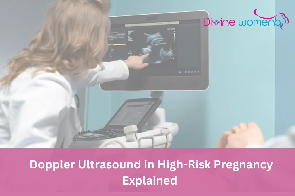
Doppler Ultrasound in High-Risk Pregnancy Explained
Pregnancy is a beautiful journey, but for some women, it may come with complications that require closer monitoring. In such cases, doppler ultrasound in high risk pregnancy plays a vital role in ensuring the health and safety of both mother and baby.
Read moreWe are here for you
Book a consultation with our women's health team in Ahmedabad
Obstetricians, gynecologists, fertility specialists and a neonatologist, with hospital staff available 24×7 for you and your baby.